Coronary Arteries Corresponding Ecg Leads / Coronary arteries of human heart and 12-standard ECG leads ... - In other instances, changes are subtle and might be recognized only when ecg recording is repeated after changes in the severity of symptoms.
Get link
Facebook
X
Pinterest
Email
Other Apps
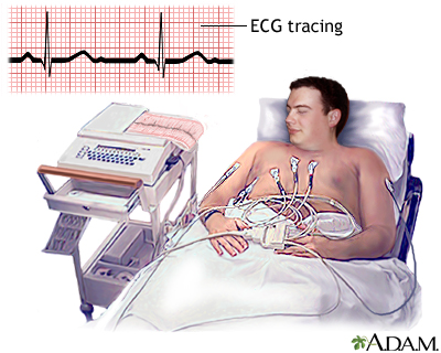
Coronary Arteries Corresponding Ecg Leads / Coronary arteries of human heart and 12-standard ECG leads ... - In other instances, changes are subtle and might be recognized only when ecg recording is repeated after changes in the severity of symptoms.. At times, the changes are typical and clear. That could cause a heart attack. Small branches dive into the heart muscle to. Coronary artery disease, is the leading cause of death in the united states for both men and women. Electrocardiography is the process of producing an electrocardiogram (ecg or ekg).it is a graph of voltage versus time of the electrical activity of the heart using electrodes placed on the skin.
In acute coronary syndromes, the electrocardiogram (ecg) provides important information about the presence, extent, and severity of myocardial ischemia. Fqrs was defined as a qrs duration of 120 milliseconds or less, with notches or slurs of qrs complexes, on 2 contiguous leads of a coronary artery territory. The coronary arteries and their relation to the ecg leads. Your ekg is recorded before, during and after the test. Spontaneous coronary artery dissection (scad) is an uncommon but dangerous condition in which one of the arteries that supply the heart spontaneously develops a blood collection, or hematoma, within the artery wall.this leads to a separation and weakening of the walls of the artery.
12-Lead STEMI| Cardiac Life from cardiaclife.net When the coronary arteries narrow to the point that blood flow to the heart muscle is limited (coronary artery disease), collateral vessels may enlarge and become active. Your ekg is recorded before, during and after the test. Second, that plaque can break open, which can lead to a blood clot. Coronary artery ectasia (cae) is an angiographic finding characterized by dilation of an arterial segment with a diameter at least 1.5 times that of its adjacent normal coronary artery. These electrodes detect the small electrical changes that are a consequence of cardiac muscle depolarization followed by repolarization during each cardiac cycle (heartbeat). It can be used to check for a number of things including: Electrocardiographic sign of coronary artery obstruction. Coronary artery disease, is the leading cause of death in the united states for both men and women.
Plaque is made up of fat, cholesterol, calcium, and
In acute coronary syndromes, the electrocardiogram (ecg) provides important information about the presence, extent, and severity of myocardial ischemia. The coronary calcium scan tells you how much calcified plaque is in your heart's arteries. Fqrs was defined as a qrs duration of 120 milliseconds or less, with notches or slurs of qrs complexes, on 2 contiguous leads of a coronary artery territory. The coronary arteries are the arterial blood vessels of coronary circulation, which transport oxygenated blood to the heart muscle. Electrocardiographic sign of coronary artery obstruction. Vascular stiffness plays an important role in the pathophysiology of cardiovascular events.1, 2 aortic pulse wave velocity (aopwv), a marker of large artery stiffness, is a strong predictor of coronary artery disease (cad),3 probably because aorta conveys pulsatility to coronary vessels. An inferior infarct on ecg (inferior myocardial infarction or inferior stemi) occurs when inferior myocardial tissue supplied by the right coronary artery (rca), is injured due to thrombosis of. Coronary artery disease develops when the major blood vessels that supply your heart become damaged or diseased. The coronary arteries supply blood, oxygen and nutrients to your heart. This allows blood to flow around the blocked artery to another artery nearby or to the same artery past the blockage, protecting the heart tissue from injury. Scad can slow or block blood flow to the heart, causing a heart attack, abnormalities in heart rhythm or sudden death. Figure 2 shows the coronary arterial territories in relation to the 17 segments of the left ventricle. When the coronary arteries narrow to the point that blood flow to the heart muscle is limited (coronary artery disease), collateral vessels may enlarge and become active.
These electrodes detect the small electrical changes that are a consequence of cardiac muscle depolarization followed by repolarization during each cardiac cycle (heartbeat). Spontaneous coronary artery dissection (scad) is an uncommon but dangerous condition in which one of the arteries that supply the heart spontaneously develops a blood collection, or hematoma, within the artery wall.this leads to a separation and weakening of the walls of the artery. Typically, an anterolateral infarct pattern with abnormal deep (>3 mm) and wide (>30 msec) q waves is observed in leads i, avl, v5, and v6, absent q waves in leads ii, iii, and avf, and poor r wave. Coronary artery disease, is the leading cause of death in the united states for both men and women. Coronary arteries supply blood to the heart muscle.
Electrocardiogram | UF Health, University of Florida Health from ufhealth.org Vascular stiffness plays an important role in the pathophysiology of cardiovascular events.1, 2 aortic pulse wave velocity (aopwv), a marker of large artery stiffness, is a strong predictor of coronary artery disease (cad),3 probably because aorta conveys pulsatility to coronary vessels. Fragmented qrs (fqrs) complexes are electrocardiographic signals which reflect altered ventricular conduction around regions of a myocardial scar and/or ischaemia. The coronary arteries supply blood, oxygen and nutrients to your heart. Electrocardiography is the process of producing an electrocardiogram (ecg or ekg).it is a graph of voltage versus time of the electrical activity of the heart using electrodes placed on the skin. If you have a coronary spasm, the doctor can see it on your ekg as well as an angiogram. New content in stemi imposters chapter includes the coronary artery anatomy, right ventricular infarction, posterior infarction, and obtaining additional leads. Fqrs was defined as a qrs duration of 120 milliseconds or less, with notches or slurs of qrs complexes, on 2 contiguous leads of a coronary artery territory. Electrocardiographic sign of coronary artery obstruction.
Your ekg is recorded before, during and after the test.
The angiogram explained the unique initial ecg findings, whereby good collateral blood supply from the left circumflex artery protected the apical left ventricular segments and corresponding ecg leads (v5 and v6) from transmural ischemia. At times, the changes are typical and clear. Spontaneous coronary artery dissection (scad) is an uncommon but dangerous condition in which one of the arteries that supply the heart spontaneously develops a blood collection, or hematoma, within the artery wall.this leads to a separation and weakening of the walls of the artery. Fqrs was defined as a qrs duration of 120 milliseconds or less, with notches or slurs of qrs complexes, on 2 contiguous leads of a coronary artery territory. The coronary arteries wrap around the entire heart. Small branches dive into the heart muscle to. These electrodes detect the small electrical changes that are a consequence of cardiac muscle depolarization followed by repolarization during each cardiac cycle (heartbeat). Scad is a major cause of heart attacks in young, otherwise healthy women without known risk factors. An inferior infarct on ecg (inferior myocardial infarction or inferior stemi) occurs when inferior myocardial tissue supplied by the right coronary artery (rca), is injured due to thrombosis of. Typically, an anterolateral infarct pattern with abnormal deep (>3 mm) and wide (>30 msec) q waves is observed in leads i, avl, v5, and v6, absent q waves in leads ii, iii, and avf, and poor r wave. This allows blood to flow around the blocked artery to another artery nearby or to the same artery past the blockage, protecting the heart tissue from injury. Coronary artery disease develops when the major blood vessels that supply your heart become damaged or diseased. If you have a coronary spasm, the doctor can see it on your ekg as well as an angiogram.
If artery blockages are present, the ekg, echocardiogram, and/or nuclear scan will show the characteristic abnormalities, enabling your doctor to make the diagnosis of coronary artery disease. New content in stemi imposters chapter includes the coronary artery anatomy, right ventricular infarction, posterior infarction, and obtaining additional leads. Spontaneous coronary artery dissection (scad) is an uncommon but dangerous condition in which one of the arteries that supply the heart spontaneously develops a blood collection, or hematoma, within the artery wall.this leads to a separation and weakening of the walls of the artery. Oronary heart disease (chd), also called. When the coronary arteries narrow to the point that blood flow to the heart muscle is limited (coronary artery disease), collateral vessels may enlarge and become active.
I'm an Emergency Physician and wanted to talk about Apple ... from upload.wikimedia.org New content in stemi imposters chapter includes the coronary artery anatomy, right ventricular infarction, posterior infarction, and obtaining additional leads. Small branches dive into the heart muscle to. The coronary arteries and their relation to the ecg leads. The coronary arteries wrap around the entire heart. Your ekg is recorded before, during and after the test. That could cause a heart attack. Oronary heart disease (chd), also called. If you have a coronary spasm, the doctor can see it on your ekg as well as an angiogram.
It can be used to check for a number of things including:
Figure 2 shows the coronary arterial territories in relation to the 17 segments of the left ventricle. The coronary arteries supply blood, oxygen and nutrients to your heart. Electrocardiography is the process of producing an electrocardiogram (ecg or ekg).it is a graph of voltage versus time of the electrical activity of the heart using electrodes placed on the skin. Plaque is made up of fat, cholesterol, calcium, and Approximately one half of patients who present with signs and symptoms of acute inferior myocardial infarction have proximal occlusion of the dominant right coronary artery (rca) and also show ecg. The ecg on presentation showed normal sinus rhythm and normal pr intervals (figure 1). Oronary heart disease (chd), also called. Spontaneous coronary artery dissection — sometimes referred to as scad — is an uncommon emergency condition that occurs when a tear forms in a blood vessel in the heart. Coronary arteries supply blood to the heart muscle. In other instances, changes are subtle and might be recognized only when ecg recording is repeated after changes in the severity of symptoms. The coronary arteries and their relation to the ecg leads. Coronary artery ectasia (cae) is an angiographic finding characterized by dilation of an arterial segment with a diameter at least 1.5 times that of its adjacent normal coronary artery. Scad is a major cause of heart attacks in young, otherwise healthy women without known risk factors.
If artery blockages are present, the ekg, echocardiogram, and/or nuclear scan will show the characteristic abnormalities, enabling your doctor to make the diagnosis of coronary artery disease coronary arteries ecg. Approximately one half of patients who present with signs and symptoms of acute inferior myocardial infarction have proximal occlusion of the dominant right coronary artery (rca) and also show ecg.


Comments
Post a Comment